Surgical Anatomy of the Liver
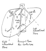
Understanding liver anatomy is of importance in liver resection. The liver lies in the abdominal cavity, where it is split into a large right and a small left lobe by the falciform ligament extending from the anterior abdominal wall. The morphology does not correspond to the surgical anatomy of the liver and functionally the liver is divided into a right and left hemi-liver by the principal plane (Rex-Cantlie line). This is a plane passing through the gallbladder bed towards the vena cava and passes through the right axis of the caudate lobe. The middle hepatic vein lies in this plane. Although this was first recognised by Ton That Tung in 1939, it was Couinaud in 1957 who provided the definitive description.
The right hemi-liver is divided into anterior and posterior sections by the course of the right hepatic vein which lies in the inter-sectional plane. This plane passes coronally through the right liver at the level of the inferior vena cava. The left hemi-liver is divided into lateral and medial sections by the left hepatic vein. Further portal inflow division results in each section in turn being subdivided into two segments with the exception of the left lateral section (one segment only). The divisions of the portal vein are mirrored by divisions of the bile duct and hepatic artery forming a 'portal trinity', a division of which supplies each segment.
The right portal pedicle is short (less than 1 cm in most) and the vein divides to supply the right anterior section, subdivided into segments V (inferior) and VIII (superior) by portal vein divisions and the right posterior section subdivided into segments VI (inferior) and VII (superior) by portal vein divisions. The left portal pedicle is long. It gives of a caudate branch and thereafter the vein divides to supply a left lateral section and a left medial section. The left medial section is divided into two segments, III and IV, by a further portal vein division. The left lateral section is the one exception to the rule as there is no further major portal vein division. Thus it only has one segment, segment II. The caudate lobe is a distinct anatomical segment and is labelled segment I. It receives branches of the portal trinity from the right and left liver and drains independently into the vena cava.
|
||
 |
||
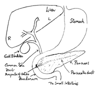
| Schematic diagram of liver, pancreas and bile ducts |
||
As each segment of the liver has its own supply from the portal trinity, independent of the other segments, these can therefore be resected (removed) independently of other segments. In practice, it is easier to remove some segments together. Although the inter-segmental planes are not visible on the surface of the liver, segments can be defined by occluding the inflow to that segment thus rendering the segment ischaemic: the change in colour at operation helps define the border.
The major hepatic veins do not correspond to the segmental division of the liver. The three named superior hepatic veins (right, middle and left) lie in the 3 main fissures and between the 4 hepatic sections. The right vein lies in the right fissure between the right anterior section and posterior section, the middle vein in the principal plane between the right and left hemi-liver, and the left vein between the left medial and lateral section. Each vein drains the section on either side of it. The right vein drains into the vena cava independently, but the middle and left veins usually join and drain into the vena cava as a single vein. There are usually a few small veins draining into the vena cava from behind the liver. Occasionally there can be 2 or 3 inferior right hepatic veins of moderate size and these can provide significant drainage. If these are not recognized and torn during hepatic resection bleeding may be profuse.
It has been recognised that Glisson's capsule extends as a condensation of fascia around the bilio-vascular branches of the portal trinity (Glissonian sheath's) Couinaud and more recently Launois and Jamieson have noted that the fascia continues within the liver parenchyma up to the segmental divisions. The surgical implication is that if the supply to an individual segment is approached from within the liver, mass ligation of a sheath will devascularise the segment. This is simplified even further by the use of a stapler.
